 Laue
Crystallography in brief
A few pages to place what Laue does. They show what a crystal is and how its symmetry is described. They then show how diffraction lets us see it, and the steps that lead from an unknown sample to the positions of its atoms.
A crystal: a lattice and a motif
A crystal is an arrangement of atoms that repeats identically in the three directions of space. It is described in two parts.
- The lattice is the set of translations that leave the crystal unchanged. It is made of points, without atoms.
- The motif is the group of atoms placed at every lattice point.
In short: crystal = lattice + motif. Three vectors a, b, c generate the lattice, and the parallelepiped they span is the unit cell. Its six parameters (a, b, c, α, β, γ) and the coordinates of the atoms, as fractions of these vectors, are enough to describe the whole crystal.
There are two kinds of cells:
- a primitive cell contains a single lattice point;
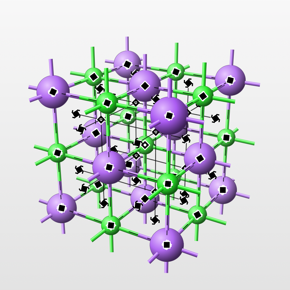
- a conventional cell is sometimes larger, but its edges follow the symmetry. The face-centred cubic cell of rock salt contains four lattice points.
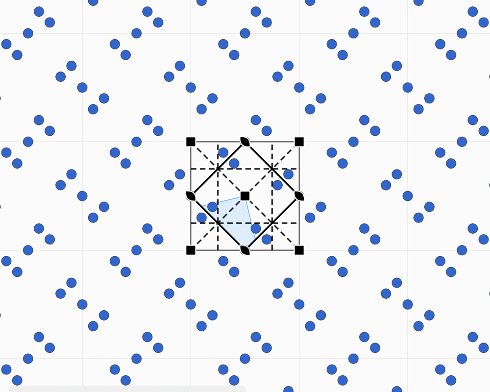
In the plane, the idea is the same with two vectors: a wallpaper, a tiled floor or a fabric are “two-dimensional crystals”.
The formulas
A lattice point, and any point in fractional coordinates:
The metric tensor holds the six cell parameters; it gives distances, angles and the volume:
Symmetry, level by level
Periodicity strongly restricts the possible symmetries. A lattice can only have rotation axes of order 1, 2, 3, 4 or 6: this is the crystallographic restriction. A regular tiling of pentagons is impossible, and so is a crystal with 5-fold symmetry. Symmetries are therefore classified by levels, each one building on the previous.
- The point group gathers the rotations, mirrors and inversion that leave a point fixed. It tells what the crystal looks like from afar, without seeing its atoms: the shape of its faces, the anisotropy of its properties. There are 32 of them. They are drawn in stereographic projection and divided into 7 crystal systems, from triclinic to cubic.
- The Bravais lattices are the 14 ways of arranging lattice points in space: primitive (P), body-centred (I), face-centred (F), one-face-centred (A, B or C), rhombohedral (R).
- The space group combines the two. It adds operations that have no point equivalent: screw axes (a rotation followed by a fraction of a translation, such as 21 or 42) and glide planes (a mirror followed by a half translation). There are 230 space groups, and 17 plane groups in the plane. Their Hermann–Mauguin symbol, such as
Fm-3morP2_1/c, gives the lattice type, then the symmetry elements along the principal directions. - Wyckoff positions classify the points of the cell by their local symmetry. An atom placed on a symmetry element has fewer copies in the cell than an atom in a general position. The product of its multiplicity and the order of its site symmetry is constant. This is the language of structure descriptions: “Na on 4a, Cl on 4b”.
| In the plane | In space | |
|---|---|---|
| Crystal systems | 4 | 7 |
| Point groups | 10 | 32 |
| Bravais lattices | 5 | 14 |
| Plane groups, space groups | 17 | 230 |
Symmetry also shows in physical properties. According to Neumann's principle, every property of a crystal has at least the symmetry of its point group. A cubic crystal is therefore optically isotropic. Piezoelectricity, on the other hand, requires the absence of a centre of symmetry.
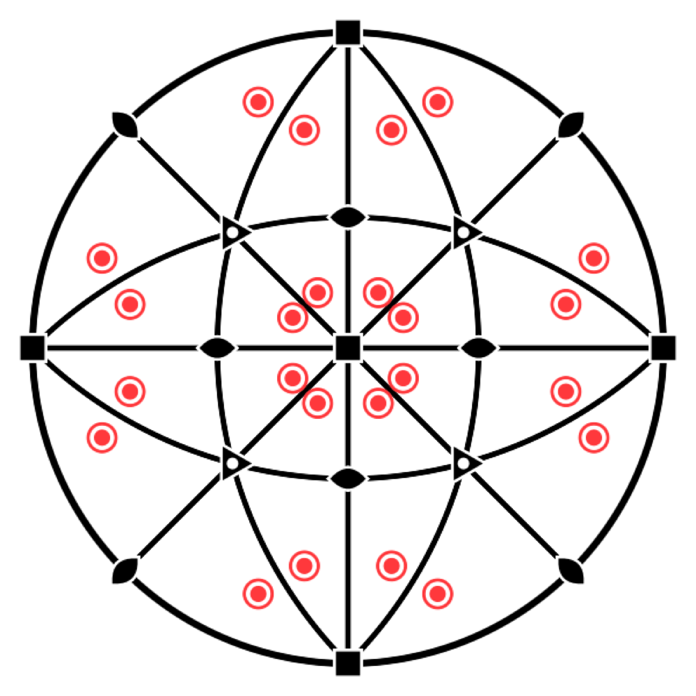
In Laue, the Tiling workshop covers the 17 plane groups and the 7 frieze groups. The Crystal workshop covers the 230 space groups, with their Wyckoff positions, their symmetry elements in the cell and their stereograms.
The formulas
A symmetry operation in Seitz notation: a linear part and a translation (fractional for a screw axis or a glide plane):
The crystallographic restriction: in a lattice basis, is an integer matrix, and so is its trace. For a rotation by :
A Wyckoff position: its multiplicity (in the conventional cell), the order of its site symmetry , the order of the point group and the number of lattice points in the cell (1 for P, 2 for I and C, 4 for F):
Seeing atoms: diffraction
Interatomic distances, of the order of an ångström (10−10 m), are far too small for an optical microscope. The crystal is therefore illuminated with a wave of comparable wavelength: X-rays, neutrons or electrons. Each atom scatters a little of the wave. Because the crystal is periodic, these waves add up only in certain directions, where they form sharp spots, the reflections. Max von Laue observed this in 1912; William Lawrence Bragg gave its law the following year.
- Where the crystal diffracts: Bragg's law. The crystal can be seen as families of parallel lattice planes, labelled by their Miller indices (h k l) and spaced dhkl apart. A family reflects the wave when 2 d sin θ = n λ. Each reflection corresponds to a point of the reciprocal lattice, at distance 1/d from the origin: the geometry of the diffraction pattern therefore gives the cell.
- With what intensity: the structure factor. The intensity of each reflection depends on the position and the nature of the atoms in the cell, through the structure factor Fhkl. It is what carries information about the motif.
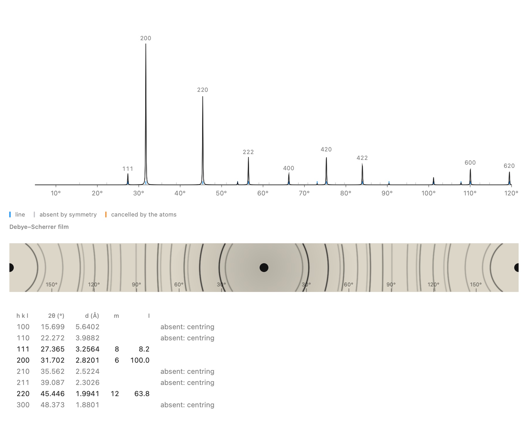
- Systematic absences. Some reflections are zero whatever the atoms. A centred lattice extinguishes whole families; a screw axis or a glide plane extinguishes others. These absences reveal the lattice type and part of the space group.
- The Laue class. The diffraction pattern is always centrosymmetric (this is Friedel's law): it shows the symmetry of the point group with a centre added, its Laue class.
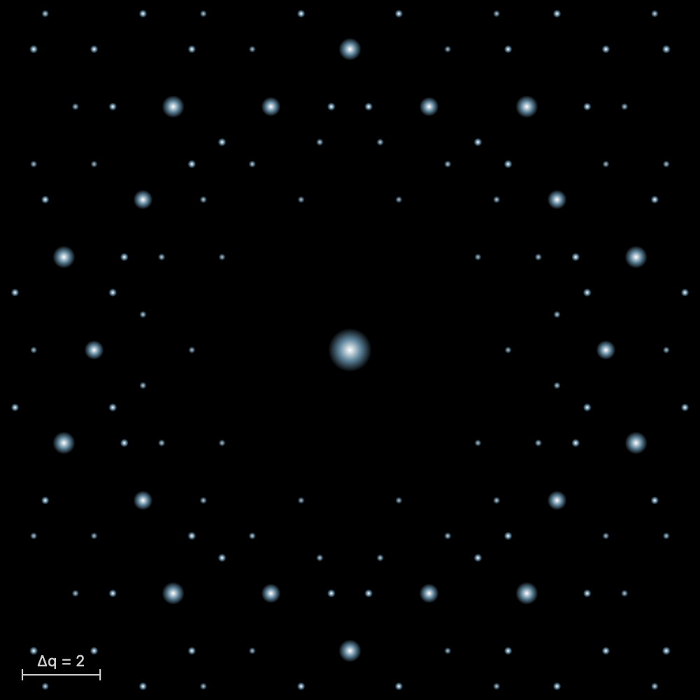
In Laue, the diffraction of a tiling, a quasicrystal or a crystal is computed with every change. You see the allowed reflections, those the group extinguishes and the reason for each absence.
The formulas
The reciprocal lattice (crystallographers' convention, without ) and the interplanar spacing:
Bragg's law, and its equivalent, the Laue condition (Ewald sphere, ):
The structure factor, a sum over the atoms of the cell (scattering factor , Debye–Waller factor ), and the intensity:
An absence: an F lattice puts an atom at and at , , , hence
Friedel's law (without anomalous scattering): , hence .
Probes and methods
Three probes complement each other:
- X-rays, scattered by electrons: the method of every laboratory, with a tube or a synchrotron;
- neutrons, scattered by nuclei. They see light atoms such as hydrogen, tell neighbours in the periodic table apart and reveal magnetic order; they need a reactor or a spallation source;
- electrons, which interact so strongly that a nanocrystal is enough, in an electron microscope.
The sample then dictates the method:
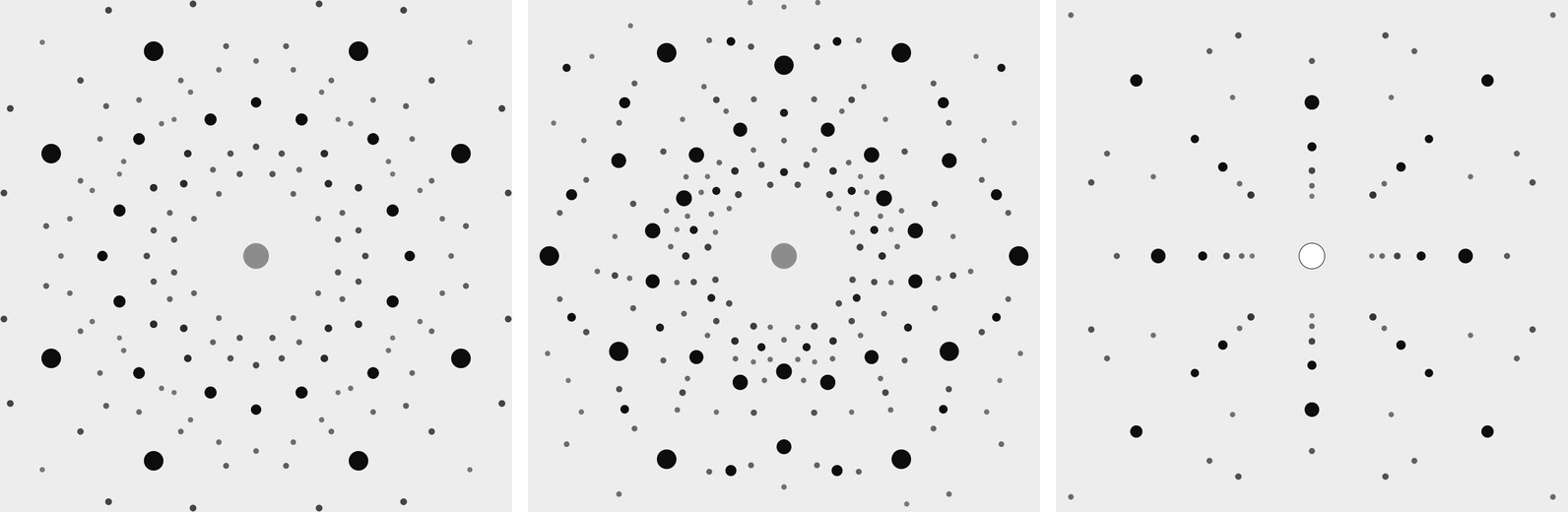
- Single crystal. The Laue method illuminates a stationary crystal with white radiation: the photograph shows its symmetry and is used to orient it. A modern diffractometer rotates the crystal in a monochromatic beam and measures thousands of reflections, one by one: this is the royal road to a complete structure.
- Powder. Thousands of randomly oriented crystallites give a diffraction cone for each family of planes. The Debye–Scherrer film shows it as arcs; a diffractometer unrolls it into a plot of intensity against 2θ. The pattern is the fingerprint of a phase. The positions of the lines give the cell, their intensities the contents of the cell, their widths the size of the crystallites (Scherrer formula).
Reading a Laue pattern. Each spot comes from a family of planes that has taken the Bragg wavelength from the white beam. The planes act as a mirror: their normal is the bisector of the angle between the beam and the ray going from the crystal to the spot. From the position of the spot on the film and the distance of the film, one therefore gets the direction of the normal, then its indices (h k l) by projecting it on the axes of the cell. The wavelength of each spot remains unknown: the pattern gives the orientation of the crystal and its Laue class, not its spacings. In Laue, the Laue pattern follows the view of the Crystal workshop, and pointing at a spot gives its indices.
The formulas
The wavelength of a neutron of velocity , and of an electron accelerated through a voltage (with the relativistic correction):
The Scherrer formula: the size of the crystallites, from the full width at half maximum of a line (in radians, instrumental broadening removed; ):
From measurement to structure
From an unknown sample to the list of atomic positions, the procedure comes down to four steps.
- Identify. The powder pattern is compared with a database of known phases. These are structure databases such as the free Crystallography Open Database, or pattern databases such as the ICDD Powder Diffraction File. Most often the substance is already known and the analysis stops there.
- Index. Otherwise, one looks for the cell that assigns indices (h k l) to every line. For a cubic cell, it is enough to see that the values of 1/d² are in the ratios of integers h² + k² + l². The absences then give the compatible space groups.
- Solve. The measurement gives the intensity of the reflections, |F|², but not their phase. Without phases, the electron density cannot be reconstructed: this is the phase problem. There are several ways around it:
- the Patterson function, which does without phases and shows the vectors between atoms;
- direct methods, which guess the phases from statistical relations (Nobel Prize 1985);
- charge flipping, which alternates between real space and reciprocal space.
- Refine and validate. The model is fitted to the data by least squares: positions, thermal motion and, for a powder, the whole profile of the pattern (Rietveld method). Agreement factors such as R and χ² tell whether the model is good.
In Laue, the Crystal workshop computes the powder pattern of a structure, with or without the real atoms. It reads a real measurement and refines it (cell, zero shift, line profile, background). It indexes an unknown measurement and proposes the compatible groups. The Fourier synthesis of tilings also shows the phase problem: with the true phases, the motif comes back; with random phases, it disappears.
The formulas
The electron density is the Fourier transform of the structure factors, each of which has a modulus and a phase ; the measurement gives only the modulus:
The Patterson function does without phases; its peaks lie at the vectors between atoms:
Rietveld refinement minimises the weighted difference between the measured pattern and the calculated pattern , point by point; the result is judged by and ( points, parameters):
Further reading
- The International Tables for Crystallography, Volume A, the reference for plane groups and space groups.
- The IUCr teaching pamphlets, short and free.
- The Bilbao Crystallographic Server, for groups, their subgroups and their Wyckoff positions.
- The Crystallography Open Database, hundreds of thousands of structures in CIF that Laue can open.
For practice, the guide describes each workshop, and the examples open in Laue with one click.